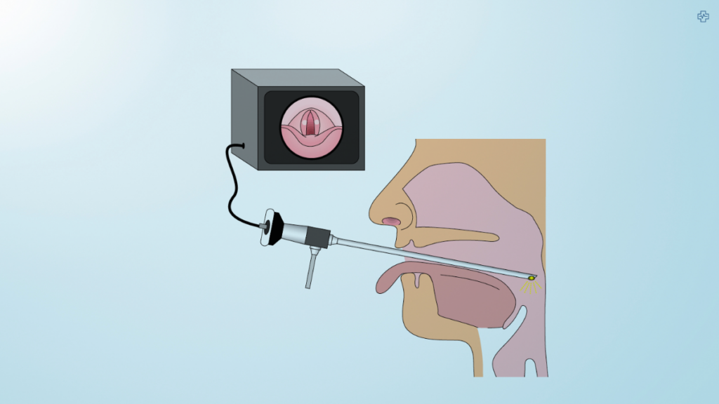
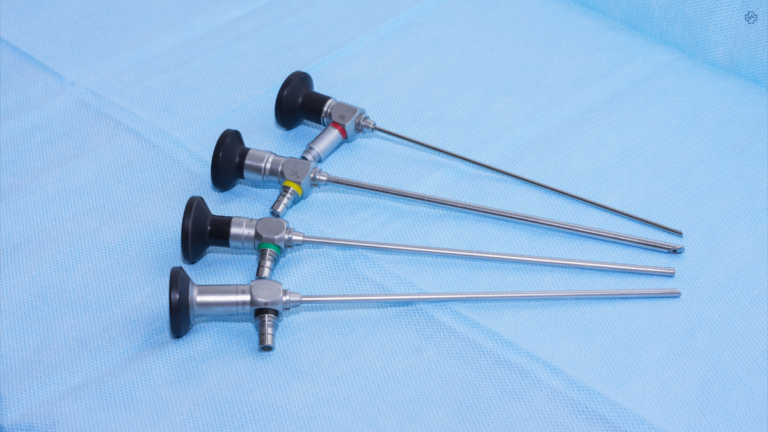
Hyperspectral imaging, or HSI,, in simple terms, is a new way to take pictures of our body that helps doctors diagnose diseases more accurately. It’s like other imaging technologies, but better. Now, scientists have made a special camera that can look at the human body in a new way, from visible light to very tiny light wavelengths. This can help doctors during surgery and make diagnosis more precise. This new invention was made by researchers at Tokyo University of Science and it’s going to change how we see and treat our bodies.
The Groundbreaking Technology of the Rigid Endoscope
Traditional imaging methods capture data at specific wavelengths, but HSI captures a full spectrum of data at every pixel, providing detailed information about the composition of tissues. This rigid endoscope, designed specifically for medical applications, integrates a supercontinuum (SC) light source with an acousto-optic tunable filter (AOTF).
This setup allows the system to capture and switch between a wide range of wavelengths within milliseconds. It can offer real-time imaging of things as delicate as blood vessels, nerves, and even cancerous tissues during surgery. The ability to image in both the visible and near-infrared (NIR) ranges enhances diagnostic accuracy, providing unprecedented clarity in the differentiation between healthy and diseased tissues.
This capability is particularly important for surgical navigation. During operations, surgeons can use this technology to visualise tumour margins more clearly. This can potentially reduce the risk of leaving behind cancerous cells or damaging critical structures. Moreover, by extending into NIR wavelengths, it enables non-invasive imaging of deeper tissues. This makes it a critical tool for procedures like cancer resections or nerve preservation during complex surgeries.
| Feature | Description |
|---|---|
| Definition | A technique that captures images across a wide range of wavelengths, beyond the visible spectrum. |
| Data Collection | Acquires hundreds or thousands of narrow spectral bands, providing detailed information about the composition of materials. |
| Applications | Agriculture, remote sensing, medical imaging, food inspection, and materials analysis. |
| Advantages | Provides rich spectral information, enabling identification of materials and detection of subtle changes. |
| Challenges | Requires specialized equipment and complex data analysis techniques. |
| Types of HSI | Airborne, satellite-based, and ground-based systems. |
| Spectral Resolution | The ability to distinguish between different wavelengths of light. |
| Spatial Resolution | The ability to resolve fine details in the image. |
| Noise | The level of random variation in the image data. |
Applications Beyond Surgery
The applications of Hyperspectral Imaging (HSI) extend far beyond the operating room, offering exciting possibilities for non-invasive diagnostics in various medical fields. In dermatology, HSI can be used to assess skin conditions, such as detecting melanoma or monitoring wound healing, by capturing detailed spectral information about tissue composition. Retinal diseases, such as age-related macular degeneration or diabetic retinopathy, can also be diagnosed early with HSI, as the technology allows for the detection of subtle changes in the retina that are not visible with traditional imaging methods.
HSI’s ability to visualise and analyse how light interacts with biological tissues can be employed to monitor the progression of internal diseases like liver fibrosis or inflammatory bowel disease without the need for invasive biopsies.
HSI’s detailed spectral data can also support pharmaceutical research by monitoring drug distribution and interaction at the cellular level. For example, the technology can track how drugs are absorbed, metabolised, and eliminated by the body, allowing for the precise mapping of drug efficacy and toxicity. This capability can revolutionise personalised medicine by enabling researchers to better understand individual responses to treatment and tailor therapies accordingly.
HSI combined with neural networks shows promising results in experimental settings, where it has already demonstrated the ability to classify different types of materials with high accuracy. The potential to detect early signs of neurodegenerative diseases like Alzheimer’s or diabetes, through non-invasive monitoring of tissue changes, could significantly improve early diagnosis and treatment outcomes.
Challenges & Future Direction

Despite its immense promise, hyperspectral imaging (HSI) technology faces several critical challenges that limit its widespread adoption. The primary challenge is the massive volume of data generated by high-resolution HSI scans, which cover hundreds of spectral bands. This immense data load requires advanced computational tools for processing and real-time analysis, posing a barrier to efficient clinical use.
Hospitals and research institutions must invest in significant IT infrastructure and machine learning algorithms to handle, process, and interpret the data effectively. Additionally, the complexity of these systems means healthcare professionals need specialised training to interpret the images and understand the results, adding another layer of complexity to its integration into regular clinical workflows.
The high cost of developing, maintaining, and operating HSI systems also limits accessibility. The components needed, such as supercontinuum light sources and advanced detectors, are expensive, and building a clinical infrastructure around these systems presents a significant financial hurdle for many healthcare providers. However, ongoing research is focused on overcoming these barriers.
Manufacturers are making efforts to miniaturise the technology making it more portable for smaller clinics and hospitals. Simultaneously, innovations aimed at reducing production costs could make these systems more affordable. If these challenges are addressed successfully, HSI has the potential to move from a specialised tool for research institutions to a widely used diagnostic technology in mainstream healthcare settings.
Industry Players Driving Innovation
Several companies are leading the charge in the HSI field, alongside research institutions. Headwall Photonics, Konica Minolta, and Norsk Elektro Optikk have been at the forefront of developing HSI systems for both medical and industrial applications. They aim to refine the technology further for broader clinical use, with a focus on improving both accuracy and accessibility. In the future, HSI could be integrated with other imaging technologies, such as PET scans or CT to offer comprehensive diagnostic solutions.
| Company | Focus |
|---|---|
| Headwall Photonics | Hyperspectral imaging cameras and sensors |
| Resonant Inc. | Hyperspectral imaging systems for remote sensing and industrial applications |
| Specim | Hyperspectral imaging cameras and solutions |
| Surface Optics Corporation | Hyperspectral imaging systems for various industries |
| Teledyne FLIR | Thermal imaging and hyperspectral imaging solutions |
| Harris Corporation | Hyperspectral imaging systems for defense and intelligence |
| Honeywell | Hyperspectral imaging sensors and systems |
| Optech | Lidar and hyperspectral imaging systems |
| Rigaku | X-ray and hyperspectral imaging instruments |
| Revenco | Hyperspectral imaging systems for agriculture and environmental monitoring |
| Konica Minolta | Hyperspectral imaging technology for material identification, food inspection, and more |
| ClydeHSI | High-performance hyperspectral imaging cameras and systems |
| CytoViva | Hyperspectral imaging for nanomaterials and biology applications |
| Pixxel Space | Satellite-based hyperspectral imaging for agriculture, environmental monitoring, and mineral detection |
| Anduril Industries | AI-enabled airborne hyperspectral imaging for defense and surveillance |
| Park Systems | Nanoscale infrared spectroscopy for chemical mapping |
| Hycroft Mining | Hyperspectral surveys for mineral exploration |
| Ceinsys Tech Limited | Geospatial technologies, including hyperspectral imaging |
| Skyfi | AI-integrated hyperspectral imagery for mineral identification |
Medical Imaging Evolution
This camera, called a rigid endoscope, can see in different ways and help doctors do surgery more precisely. It’s also super helpful for figuring out what’s wrong with you quickly and accurately. As this tech gets better and more doctors use it, it could make healthcare a whole lot better and more accurate. Plus, with the medical imaging industry expected to be worth over £35 billion by 2025, this kind of camera is going to be a big part of the future of medicine.

