
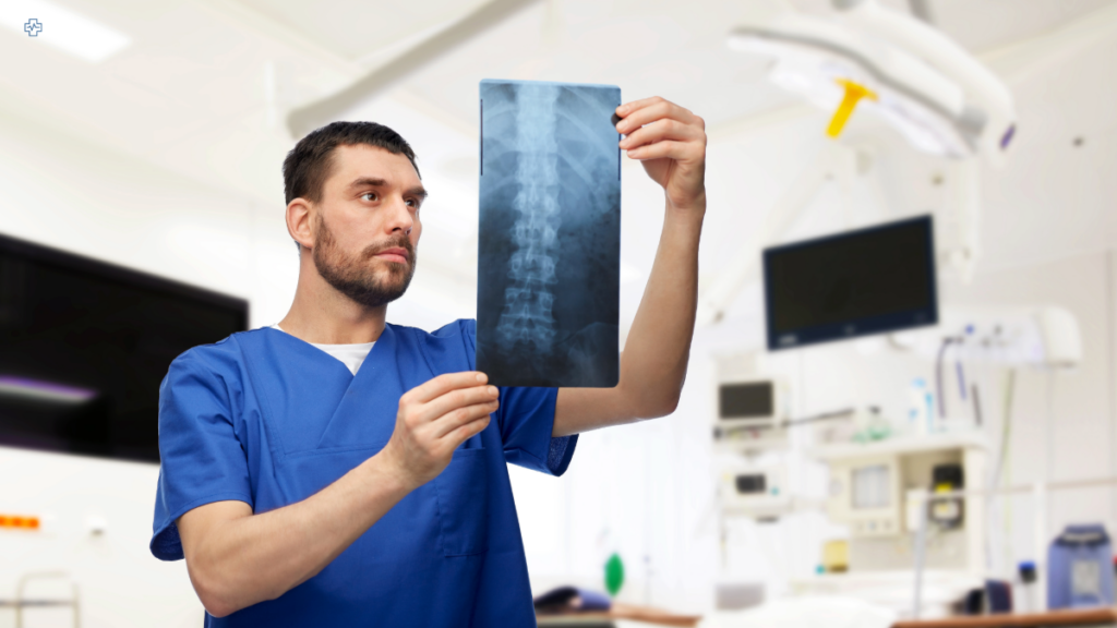
In the late 19th century, a German physicist, Wilhelm Röntgen, was conducting experiments on electrical discharges through vacuum tubes. On a cold November night in 1895, he made a world-changing discovery. As Röntgen passed electricity through the tubes, he noticed that a fluorescent screen in his laboratory began to glow. Despite no direct light source. Intrigued, he continued his experiments, eventually producing what we now know as the first X-ray image. A shadowy portrait of his wife’s hand, complete with rings and bones. This accidental discovery opened the door to a new era of medical imaging. One that would forever change healthcare.
Fast forward to 1913, and another breakthrough occurred. Albert Salomon, a German surgeon, was working with X-rays, curious to explore how they could be used to study breast tissue. Salomon’s work became the first recorded instance of what we now call a mammogram. Though rudimentary by today’s standards, Salomon’s efforts laid the foundation for the critical role mammography would eventually play in detecting breast cancer early. A disease that, at the time, was often fatal due to late-stage detection.
Salomon’s pioneering work was just the beginning. In 1927, Otto Kleinschmidt took a significant step forward by capturing the first X-ray image of breast tissue from a living patient. This was a pivotal moment in medical imaging, demonstrating the real-world potential of mammography. But true progress was slow, and it wasn’t until 1930 that Stafford Warren, an American radiologist, advanced the practice even further. Using stereoscopic X-rays, Warren studied breast tissue changes over time, foreshadowing the future of diagnostic imaging. At this point, mammography was still in its infancy, more experimental than reliable.
The 1960s, however, marked a turning point. Dedicated mammography machines were developed with enhanced image quality, offering clearer, more detailed views of breast tissue. These machines were the first designed specifically to detect breast abnormalities, distinguishing them from the general-purpose X-ray devices used previously. For women, this meant the possibility of earlier detection, which, in the fight against breast cancer, could make all the difference between life and death.
By the 1970s, the medical community began to fully grasp mammography’s potential. Large-scale studies demonstrated the effectiveness of mammograms in detecting breast cancer at an earlier, more treatable stage. These studies spurred health authorities into action. In 1976, the American Cancer Society took a bold step, recommending that women over 40 should undergo annual mammograms. This move marked a pivotal moment in breast cancer awareness and prevention, solidifying mammography’s role in saving lives.
Technological advancements continued to push the boundaries of what was possible. By the 1990s, digital mammography had emerged, offering clearer images and reducing the need for multiple views – a game-changer in terms of both comfort and accuracy for patients. No longer reliant on film, digital mammography allowed radiologists to enhance and manipulate images for better diagnosis. Women undergoing screening could now expect more accurate results, fewer callbacks, and a quicker, more efficient process.
Mammography Timeline
| Year | Event |
|---|---|
| 1895 | Wilhelm Röntgen discovers X-rays. |
| 1913 | Albert Salomon performs the first mammograms. |
| 1927 | Otto Kleinschmidt publishes the first known image of breast tissue. |
| 1930 | Stafford Warren uses stereoscopic X-rays to study breast tissue. |
| 1960s | Dedicated mammography machines are developed. |
| 1970s | Studies demonstrate the effectiveness of mammography. |
| 1976 | American Cancer Society recommends annual mammograms. |
| 1990s | Digital mammography becomes more widely available. |
| 2000s | 3D mammography is introduced. |
| 2010s | AI improves mammography image analysis. |
The turn of the millennium saw another breakthrough: 3D mammography, or tomosynthesis. Introduced in the 2000s, this technology revolutionised how breast tissue was examined. Instead of relying on two-dimensional images, 3D mammography allowed radiologists to view multiple layers of breast tissue, providing a more detailed and nuanced picture. This was especially helpful in detecting smaller or hidden tumours that might have been missed by traditional methods.
In the 2010s, the power of artificial intelligence began to make its mark on mammography. AI systems, equipped with machine learning algorithms, began analysing mammogram images to detect abnormalities with remarkable accuracy. These systems can spot patterns invisible to the human eye, offering a second layer of analysis to support radiologists. Today, AI-assisted mammograms are helping doctors detect cancer earlier, improving outcomes for thousands of women each year. It is a far cry from Albert Salomon’s early experiments, yet the core mission remains unchanged: to detect breast cancer as early as possible and give patients the best chance of survival.
As we look back on the evolution of mammography, it is remarkable to see how far we have come. What started with Röntgen’s accidental X-ray discovery over a century ago has now become an indispensable tool in women’s healthcare. But mammography is not the only medical imaging technology to undergo such a profound transformation. Similar advancements have occurred in the fields of ultrasound, MRI, and CT scanning, each playing a critical role in diagnosing and treating conditions that were once untreatable or detected too late.
The Latest Tech
Today, mammography is at the cutting edge of medical technology, with AI and machine learning poised to play an even larger role in the future. AI has already shown its potential to improve detection rates and reduce false positives, but it is just the beginning. Researchers are exploring how AI might one day predict breast cancer risk, offering personalised screening plans based on an individual’s unique risk profile. Imagine a future where mammograms are tailored to a patient’s genetics, lifestyle, and history, offering a far more precise approach to screening and prevention.
However, as promising as this technology is, it is important to acknowledge its current limitations. AI, while powerful, is not infallible. It requires vast amounts of data to learn and improve and its efficacy depends on the quality of that data. There are also ethical considerations regarding privacy and the potential for AI systems to reinforce biases present in historical medical data. As we embrace these innovations, it is crucial to strike a balance between technology and human expertise, ensuring that AI remains a tool to support, rather than replace, skilled radiologists.
The story of mammography is one of relentless innovation, driven by a singular purpose: to save lives. From the first X-rays to today’s AI-enhanced imaging, mammography has evolved in ways that its early pioneers could scarcely have imagined. And yet, there is still more to come. As technology continues to advance, so too will our ability to detect breast cancer earlier and more accurately, offering hope to countless women around the world. In this ongoing journey, mammography stands as a testament to the power of science, technology, and human determination.