
Retinal imaging is emerging as a non-invasive method for detecting early signs of systemic diseases. Retinal image has the potential to pick up conditions like diabetes, hypertension, cardiovascular disease, and even Alzheimer’s disease. “Retinal imaging allows eye doctors to see signs of eye diseases that they couldn’t see before. The test itself is painless, and the results are easy for doctors to interpret,” says WebMD.
The advancement of retinal imaging techniques positions ophthalmology at the forefront of preventive medicine. This means that routine eye exams could become integral to broader health assessments.
“New imaging technologies like artificial intelligence and deep learning systems show a potential to screen populations at risk of retinal diseases at a large scale in a resource constrained setting,” says the National Library Of Medicine.
Statistics show that 10 percent of outpatient appointments in the UK are related to eye problems, integrating retinal imaging into routine screenings can facilitate early detection and hence, early treatment of certain conditions.

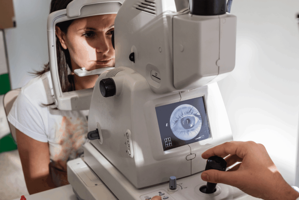
The journey of retinal imaging began in 1851 with the invention of the ophthalmoscope by Hermann von Helmholtz. This allowed physicians to view the interior of the eye for the first time.
This breakthrough paved the way for more detailed examinations of the retina. In 1926, the development of the fundus camera enabled the first photographic documentation of the retina, providing a permanent record of ocular health.
Over the decades, technological advancements have led to the creation of wide-field imaging systems and, more recently, optical coherence tomography (OCT).
Introduced in the late 1990s, OCT offers high-resolution, cross-sectional images of the retina, facilitating the detection of subtle changes which may indicate systemic diseases.
The Retina as a Mirror to Systemic Health
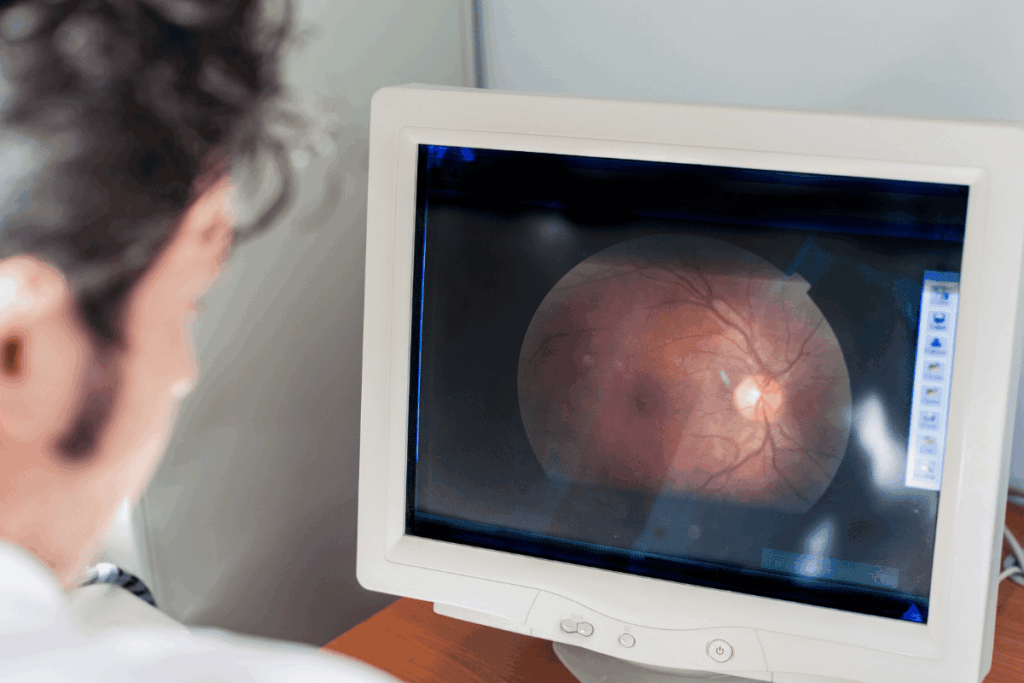
The retina has a unique position. It is part of the central nervous system and richly vascularised. This makes it an ideal site for observing systemic health. Changes in retinal morphology can reflect alterations in blood vessels and neural tissue. These nuances can indicate conditions affecting other parts of the body.
For instance, research has shown that thinning of specific retinal layers is associated with an increased risk of developing neuropsychiatric, cardiac, pulmonary, metabolic, and lung diseases.
The integration of artificial intelligence (AI) into retinal imaging has further expanded its diagnostic capabilities. Machine learning algorithms can analyse retinal images to identify patterns and biomarkers indicative of systemic diseases.
A study utilising data from the UK Biobank demonstrated that AI models could predict the risk of cardiovascular events by analysing features in retinal OCT images, achieving accuracy levels comparable to traditional risk assessment tools.
Here are 10 interesting facts about retinal imaging:
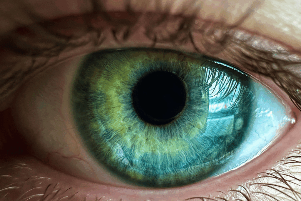
1 The retina is the only place doctors can see blood vessels directly
Unlike other parts of the body where vessels are hidden, the eye’s transparent structures let clinicians look straight at tiny arteries and veins without surgery.
2 Retinal scans can reveal systemic diseases
Conditions such as diabetes, high blood pressure, Alzheimer’s, Parkinson’s, and even heart disease often leave early clues in the retina. That’s why it’s sometimes called a ‘window to the body.’
3 AI is transforming retinal imaging
Artificial intelligence systems can now analyse retinal photographs to detect diabetic retinopathy, glaucoma, and even cardiovascular risk faster and sometimes more accurately than human specialists.
4. NASA uses retinal imaging in space
Astronauts have their retinas scanned aboard the International Space Station to monitor vision changes caused by microgravity, a condition known as Spaceflight-Associated Neuro-ocular Syndrome (SANS).
5 It can spot silent strokes
Microvascular changes in the retina can mirror those in the brain, making retinal imaging a potential tool for detecting stroke risk before symptoms appear.
6 Portable retinal cameras are making eye care accessible
Handheld, smartphone-based fundus cameras are being used in rural clinics and pharmacies, expanding access to early detection of blindness-causing conditions.
7 Retinal imaging can track biological ageing
Researchers are studying how retinal vessel changes and layer thinning correlate with ageing, potentially offering a non-invasive ‘ageing clock.’
8 The technology is centuries old but reinvented
The first fundus camera was invented in the early 1900s, but today’s devices use high-resolution digital imaging, optical coherence tomography (OCT), and even hyperspectral imaging for unmatched detail.
9 It’s painless and non-invasive
Unlike blood tests or biopsies, retinal imaging just requires a photograph or scan. This is often completed in a few seconds.
10 OCT is like an ‘optical ultrasound’
Optical coherence tomography uses light waves (instead of sound) to create cross-sectional images of the retina at near-microscopic resolution. This allows doctors to see layers thinner than a strand of hair.
Retinal imaging is proving itself to be far more than a tool for spotting eye disease. This is because the boundaries between ophthalmology and systemic medicine are starting to blur. As mentioned, this holds great promise for the world of diagnostics.
The retina is becoming a powerful diagnostic frontier, allowing for the prediction of a myriad of conditions, from cardiovascular weaknesses to neurodegenerative disorders.
The methods for this form of diagnosis are painless, and non-invasive, since the retina is easily accessible.
With advances in AI, portable devices, and next-generation imaging like OCT and hyperspectral analysis, retinal scans could soon be as routine as a blood test. Experts predict that this form of imaging will increasingly help clinicians to catch conditions earlier, thus improving treatment outcomes.