
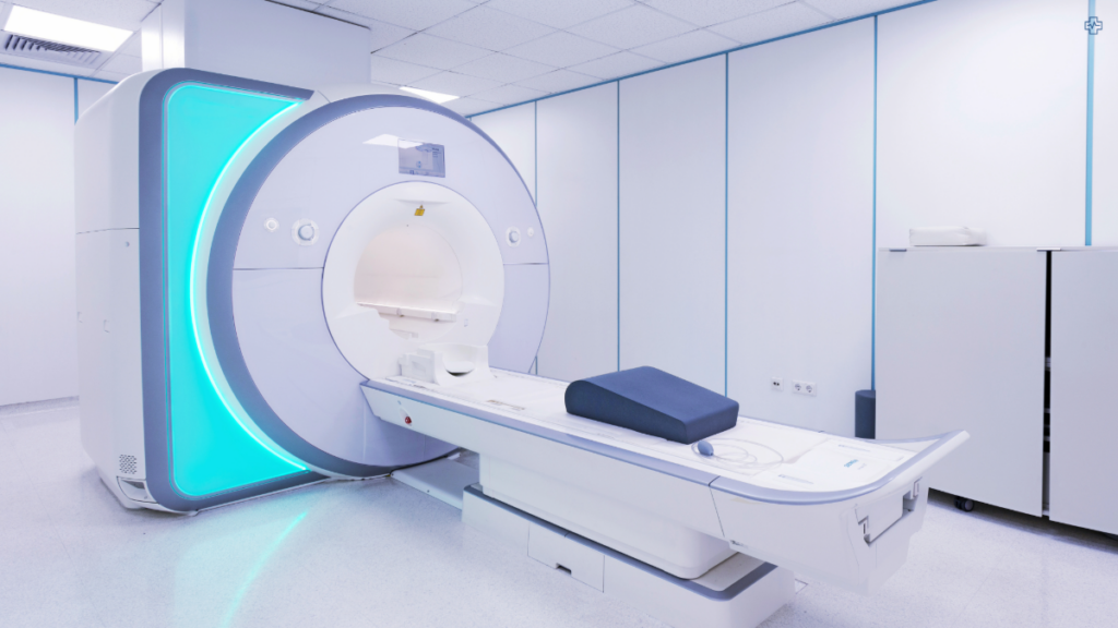
It is of little surprise that MRI technology has been making steady advances across various fields of medicine. From cardiac imaging to neurological studies MRI is increasingly used to improve detection and patient outcomes. Particularly in oncology, these innovations have opened new possibilities in the diagnosis and treatment of cancers, including prostate cancer.
A key development is the combination of multiparametric MRI (mpMRI) and Prostate-Specific Membrane Antigen (PSMA) PET scanning, which provides a more precise and detailed view of prostate cancer than ever before. This hybrid approach blends anatomical and functional imaging, offering critical insights that significantly improve diagnostic accuracy.
The Role of Multiparametric MRI & PSMA PET in Prostate Cancer
Multiparametric MRI (mpMRI) has become the cornerstone of prostate cancer diagnostics. Unlike traditional imaging methods, mpMRI combines different MRI sequences, such as T2-weighted imaging, diffusion-weighted imaging (DWI), and dynamic contrast-enhanced (DCE) MRI, to offer a comprehensive picture of the prostate. This technique can more accurately distinguish between healthy and cancerous tissue, reducing the need for invasive procedures like random biopsies.
On the other hand, PSMA PET imaging uses a radioactive tracer that binds to the PSMA protein, which is highly expressed on prostate cancer cells. The tracer highlights areas of concern, providing metabolic information that mpMRI alone cannot capture. When combined, these technologies allow for better-targeted biopsies, more effective treatment planning, and the potential to avoid unnecessary procedures in men with non-aggressive cancers.
Early studies show promising results. For example, a 2021 study found that combining PSMA PET with mpMRI improved the sensitivity for detecting prostate cancer to 97%, a significant increase compared to mpMRI alone. This combined approach also enhances staging and helps in determining whether cancer has spread beyond the prostate.
Multiparametric MRI (mpMRI) is like a super camera for doctors to take really clear pictures of the prostate to see if there’s any cancer. It uses different types of “lenses” to look inside, making it easier to spot bad (cancerous) cells and avoid random tests that might not be needed.
Benefits of the Combined Approach
By combining the detailed anatomical images from multiparametric MRI (mpMRI) with the functional data from PSMA PET, clinicians can differentiate between benign (non-cancerous) and malignant (cancerous) tissues with far greater precision. The anatomical detail provided by mpMRI, which highlights the structure of the prostate, paired with the metabolic activity detected by PSMA PET, gives a more complete picture. This synergy reduces the risk of false positives, where non-cancerous areas might otherwise be mistaken for cancer, significantly boosting diagnostic confidence and ensuring that patients receive more accurate assessments.
The hybrid imaging also allows for targeted biopsies, where tissue samples are taken specifically from areas of concern, as identified by the combined imaging results. This reduces the need for random sampling and increases the likelihood of detecting clinically significant cancers. As a result, doctors can focus on the most suspicious areas while minimising unnecessary biopsies, reducing both the discomfort and risks associated with excessive tissue sampling.
The combo of these imaging techniques aids in advanced staging of prostate cancer. Staging, which is essential for determining the extent of cancer and planning treatment, becomes more precise as the hybrid method helps detect whether the cancer has metastasized (spread beyond the prostate). This detailed information is critical in deciding whether surgery, radiation, or other interventions are necessary, ultimately guiding personalised and more effective treatment plans.
Applications Beyond Diagnosis
The combination of PSMA PET and MRI is not only a game-changer for diagnosing prostate cancer initially, but it also plays a critical role in monitoring patients after treatment. For men who have undergone prostate cancer therapies, such as surgery or radiation, these advanced imaging techniques can accurately detect whether the cancer has returned, even at very low levels. This precise detection is vital for guiding the next steps in treatment, whether that involves further surgery, hormone therapy, or radiation. By offering a detailed view of both the prostate’s structure and the metabolic activity of potential cancer cells, doctors can make more informed decisions and reduce unnecessary interventions.
The use of PSMA PET is expanding beyond prostate cancer. It is being explored for applications in detecting other types of cancers, including renal (kidney) and thyroid cancers, due to its ability to target specific cancer markers. This versatility makes PSMA PET a promising tool in oncology, as it could potentially offer more accurate diagnostics and treatment plans for a broader range of cancers. As research continues, its application could revolutionise how various cancers are managed, providing a more targeted approach to identifying and treating tumours across different organs.
MRI Machine Manufacturers
| Company | Country | Known for |
|---|---|---|
| Siemens Healthineers | Germany | Wide range of MRI machines, including high-field and ultra-high-field systems. |
| GE Healthcare | United States | Offers a comprehensive portfolio of MRI machines, with a focus on innovation and patient comfort. |
| Philips Healthcare | Netherlands | Known for its advanced imaging technologies, including MRI and PET/MRI systems. |
| Canon Medical Systems | Japan | Provides a variety of MRI machines, including open-bore systems for claustrophobic patients. |
| Toshiba Medical Systems | Japan | Offers a range of MRI machines, including high-field and ultra-high-field systems. |
| Esaote | Italy | Provides MRI systems for various clinical applications, including cardiology, oncology, and neurology. |
| Hitachi Medical Systems | Japan | Offers a variety of MRI machines, including high-field and ultra-high-field systems. |
| Mindray Medical | China | Provides a range of imaging equipment, including MRI machines. |
| Medtronic | United States | Offers a variety of medical devices, including MRI-compatible surgical tools and implants. |
Fresh Challenge
While the combination of PSMA PET and MRI offers substantial benefits, there are challenges in making this technology widely available. The cost of both PSMA tracers and advanced MRI equipment is high, and specialised training is required to interpret the images accurately. Companies like Siemens Healthineers, GE Healthcare, and Philips are investing in making MRI technology more accessible, and further developments may help bring costs down over time. Additionally, continued research is required to solidify PSMA PET/MRI as the standard of care for prostate cancer, though early results are highly encouraging.
As more clinical trials are conducted and the technology becomes more refined, it is likely that PSMA PET/MRI will play a larger role in the comprehensive management of prostate cancer, improving patient outcomes through more accurate diagnosis, staging, and treatment planning.