
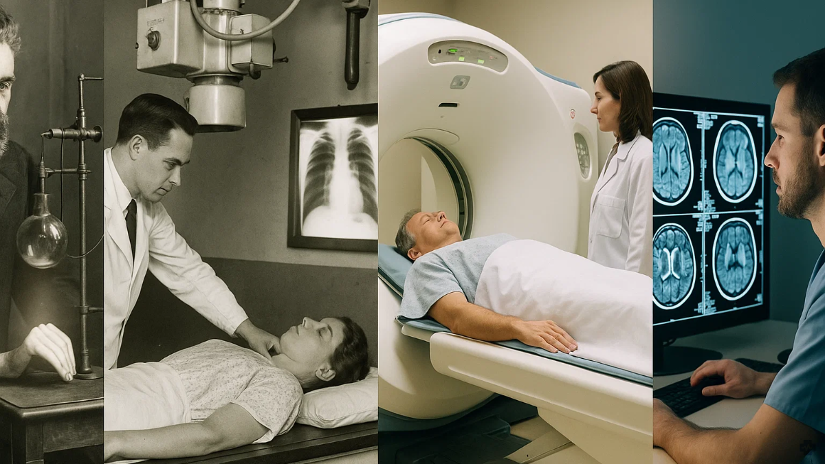
Few fields in medicine have undergone such radical transformation as radiology. What began over a century ago as the marvel of seeing inside the human body without making a single incision has evolved into one of the most technologically advanced domains in healthcare.
Radiology, once limited to rudimentary black-and-white X-ray films, now sits at the nexus of data science, digital imaging and artificial intelligence. The journey from early Roentgen rays to intelligent algorithms mirrors the broader evolution of modern medicine. Marked by relentless progress, shifting paradigms, and ever-growing expectations for precision and efficiency.
In today’s data-driven world, diagnostic imaging is no longer solely the art of interpreting shadows and densities. It is a computational science, fuelled by increasingly sophisticated tools capable of processing and analysing visual information at a scale and speed far beyond human capacity. The arrival of artificial intelligence into the radiology suite represents another step in technological advancement, a fundamental shift this time, in how clinicians approach diagnosis, triage and treatment planning.
Humble Beginnings
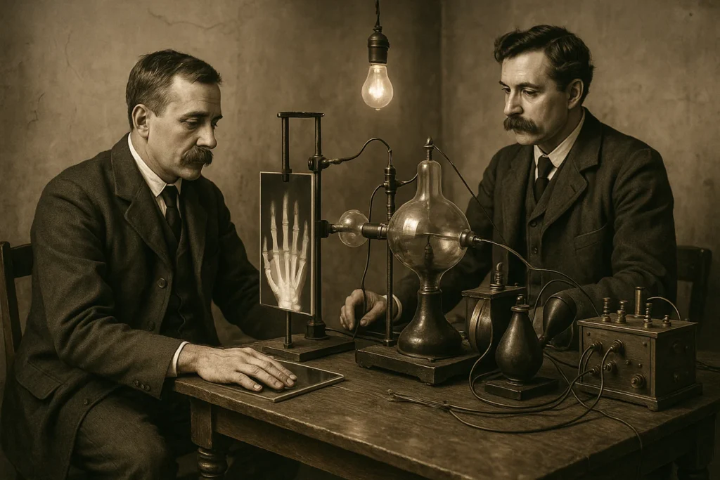
The field of radiology traces its origins to one of the most pivotal moments in medical history: the accidental discovery of X-rays in 1895 by German physicist Wilhelm Conrad Roentgen. While he was experimenting with cathode rays, Roentgen observed that a fluorescent screen in his laboratory began to glow despite being shielded by heavy black cardboard. Further investigation revealed that an invisible form of energy. What he would end up calling “X-rays”, could pass through solid objects and produce shadowy images of internal structures. His iconic first X-ray image, of his wife’s hand showing clearly defined bones and her wedding ring, stunned the medical world and laid the foundation for diagnostic imaging.
Roentgen’s discovery immediately revolutionised medical practice by enabling physicians to see inside the body without cutting it open. In the early 20th century, X-rays became standard in hospitals for diagnosing fractures, locating foreign objects and detecting diseases such as tuberculosis. However, this early form of radiology had significant limitations. Imaging was static, resolution was poor and exposure to high levels of radiation posed risks that were not initially understood.
By the 1920s and 30s, the field began to evolve with the introduction of fluoroscopy, a technique that allowed real-time imaging of moving bodily structures. This was particularly useful in gastrointestinal studies and interventional procedures. The development of contrast media (such as barium and iodine-based compounds) further expanded radiology’s diagnostic potential by enhancing visibility of soft tissues and blood vessels.
A major leap occurred in the 1970s with the invention of Computed Tomography (CT) by British engineer Sir Godfrey Hounsfield and South African physicist Allan Cormack. CT scans combined X-ray technology with computer algorithms to create cross-sectional images of the body, offering unprecedented detail. These were followed by Magnetic Resonance Imaging (MRI) in the late 1970s and early 80s, which used powerful magnets and radio waves to generate highly detailed images of soft tissue, the brain, spinal cord, and joints. And it could do this all without the risks associated with radiation.
Each of these milestones added layers of sophistication, expanding radiology beyond simple bone imaging to encompass neurological, cardiovascular, musculoskeletal and oncological diagnostics. Ultrasound, meanwhile, emerged as a widely used non-invasive modality that offered real-time imaging with no radiation exposure, especially valuable in obstetrics, cardiology, and emergency medicine.
In the 1990s, a shift from analogue to digital imaging, helped by the rise of computing power, transformed radiology into a more agile and data-driven discipline. Digital storage (PACS), faster image acquisition, and enhanced manipulation tools improved image quality and facilitated remote diagnostics and teleradiology. The transition to picture archiving and communication systems (PACS) allowed for rapid sharing of images across healthcare networks, helping to gather unprecedented data.
By the early 2000s, machine learning algorithms began entering the picture, although their utility was initially limited by processing capabilities and data availability. It wasn’t until the 2010s, with the explosion of big data and the rise of artificial intelligence (AI), that radiology once again stood at the brink of a radical transformation. AI offered the tantalising promise of pattern recognition, anomaly detection, and image interpretation at scales and speeds no human could match. This started what many now call the fourth era of radiology.
AI in Radiology
AI’s integration into radiology is a major milestone due to its profound potential to reshape diagnostic imaging.
Traditionally, radiologists must manually review images, a process that can be time-consuming and prone to error, especially when dealing with large volumes of cases. AI enhances this process by automating the detection of abnormalities, streamlining workflows, and reducing the likelihood of misdiagnoses.
By learning from vast datasets, AI tools can continuously improve in accuracy, providing more reliable and consistent diagnostic results. This advancement not only speeds up the diagnostic process but also improves the quality of care patients receive.
As such, the introduction of AI in radiology is reshaping traditional diagnostic procedures. In the past, diagnosing conditions like stroke, cancer, or fractures from medical images required radiologists to manually examine each image, a process that could take several hours.
With AI-powered tools like Viz.ai, this procedure is expedited. The AI can instantly analyse CT scans and alert healthcare professionals about critical issues, drastically reducing wait times. Furthermore, AI can assist in pre-screening, flagging potentially concerning images for further review, thus allowing radiologists to prioritise urgent cases and improve overall efficiency.
One such AI radiology application is Viz LVO, developed by Viz.ai. This is an AI-based software designed to analyse CT scan images for detecting large vessel occlusion (LVO) strokes, a severe type of ischemic stroke.
The software automatically alerts clinicians when it detects signs of this condition, allowing for faster decision-making and more timely intervention. By using AI, Viz.ai’s platform seeks to reduce the time it takes to identify critical conditions, improving patient outcomes and optimising the efficiency of healthcare delivery.
The Future of Radiology
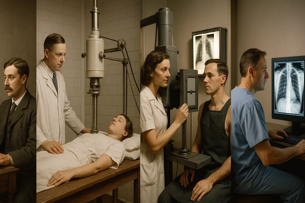
Future advancements in radiology technology are expected to be driven by continued innovations in artificial intelligence (AI), machine learning, and advanced imaging techniques. AI will likely become even more integrated into clinical workflows, providing real-time, highly accurate interpretations of medical images and enabling earlier detection of diseases like cancer, heart disease, and neurological disorders.
Personalised imaging, powered by AI, could allow for tailored diagnostic approaches based on a patient’s unique genetic make-up, leading to more precise and effective treatments. Additionally, advancements in 3D imaging, augmented reality (AR), and quantum computing could further enhance diagnostic capabilities, providing more detailed and dynamic images.
The use of robotic surgery, used in conjunction with advanced imaging, is also expected to improve the precision of minimally invasive procedures. As these technologies evolve, they will likely improve diagnostic speed, accuracy, and accessibility, while also reducing costs and enhancing patient outcomes.
Radiology has always been a field defined by innovation. From the discovery of X-rays to the integration of artificial intelligence and beyond, its trajectory reflects the broader story of modern medicine… relentless in its pursuit of better, faster, and more effective care.
As we move into an era of intelligent imaging and personalised diagnostics, radiology will not only remain a central pillar of healthcare but also serve as a blueprint for how technology and medicine can evolve together to transform patient outcomes.
The face of radiology is changing, but its mission remains the same. This is to see more clearly, so we can heal more effectively.